Body Diastasis Recti - Abdominal Muscle Separation: Case 1 (21948)
Description
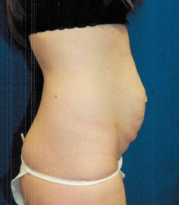
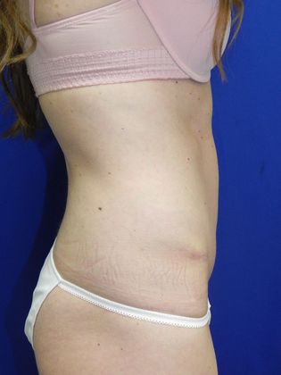
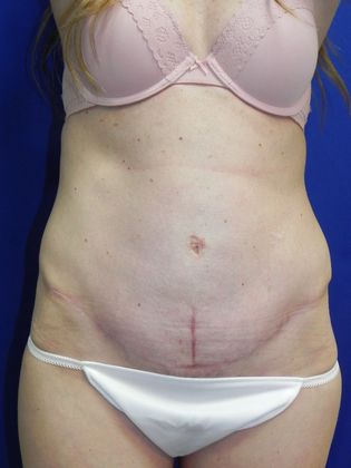
This 35 year old mom of three sought our help to repair her prominent abdominal muscle separation, known medically as diastasis recti. Her first pregnancy gave her twins. Her pregnancies were otherwise unremarkable and her youngest was now 3 years old. She works in the medical field and had heard of Dr. Graham's fine reputation. She was in good general health. She had minor scoliosis and her left hip appeared more prominent. She had a low "bikini cut" scar from Cesarean section delivery. The laxity of her abdominal wall was very visible and rather remarkable. Her other tissues were less effected: the skin looseness was relatively minor and there was no excess fat under the skin or within the abdomen. The plan was to concentrate on removing slack from the abdominal wall fibrous tissue layers (aponeurosis). We discussed measures to minimize her postop pain and speed her recovery including using Exparel (https://www.exparel.com/) which is not routinely available at area hospitals. By 4 months postop she reported doing planks, crunches and training for a marathon. She looks so much better. Viewers will notice that the postop photos show a significant vertical scar in the lower abdomen. This is the remnant of the old bellybutton site shifted downward, which would be much less noticeable if she actually had looser tummy skin. When it completely fades it will be less noticeable. The improvement in her tummy contour is excellent.
Patient Profile
- Age
- 35
- Height
- 5' 2"
- Weight
- 128
- Previous Pregnancies
- 2, singlet plus twins
- Reason for Undergoing Treatment
- Diastasis recti
- Surgical Technique
- Abdominoplasty, diastasis / muscle repair


